Three-inch wood fragment discovered in head of American quarter horse mare
October 11, 2021

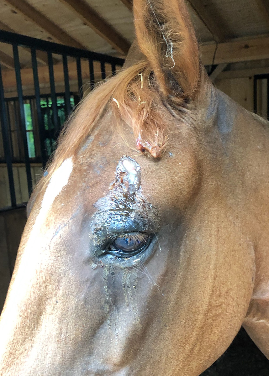
At the end of September, Love, a 16-year-old American quarter horse mare, visited the Equine Medical Center for evaluation of swelling above her left eye. The swelling had developed over several weeks, and mucopurulent discharge eventually began to ooze from the area.
To evaluate the swelling, Love’s owner Jennifer Jinnett, of Mineral, Virginia, called her primary care veterinarian, a 2013 graduate of the Virginia-Maryland College of Veterinary Medicine, Katie Huffman of Little Hawk Equine in Crozier, Virginia.
Huffman took X-rays that revealed swelling in the left temporomandibular area. After Love was treated for several days on the farm, her appetite decreased, and she started quidding - dropping food from her mouth - when attempting to eat.
Huffman decided to refer Love to James Brown, clinical associate professor of equine surgery, for a second opinion. Brown recommended a standing head CT scan to identify the soft tissue swelling over the left temporomandibular joint.

Jennifer had initially thought that Love had been stung by something. The injury had begun as a small scab and swollen area on the mare’s head, which initially started to resolve. But after four days, the infection erupted through Love’s skin just away from her eye.
Upon arrival at the Equine Medical Center, Love showed signs of pain during palpation of the affected region and resented lateral movement of her mandible. Careful examination and head CT images were consistent with left temporomandibular joint sepsis with osteolysis of the bone comprising the joint. To resolve the infection, surgical excision of the infected bone and meniscus was recommended.
Learning that her 16-year-old mare -- whom she has had since birth -- needed immediate surgery was incredibly difficult for Jennifer.
Placing Love under general anesthesia, Brown incised the area and dissected to the level of the temporomandibular articulation. Upon his entering the temporomandibular joint, a large amount of thick, viscous yellow pus flowed. A swab of fluid was submitted for Microgen next-generation sequencing.
During exploration of the area, a 3-inch piece of wood was located within the tract, extending from dorsal to the left eye into the temporomandibular joint. The wood fragment was removed, the tract was debrided, and a Jackson-Pratt drain was placed before the area was closed. Immediately before and after surgery, Love was started on intravenous antibiotics.
Within 24 hours of surgery, Love showed improvement in her comfort while chewing, and her quidding ceased. She was switched to oral antibiotics and was ready to be discharged with detailed instructions for continued medical treatment and care at home.
“Everyone was super-supportive while I fell apart in your waiting room,” shared Jennifer. “She is my world, and anyone who knows me knows I wasn't going to give up on her without a fight, so we did the surgery. And I am so glad we did! Dr. Brown and the staff at the Equine Medical Center did an extraordinary job saving my girl. Everyone there knew how much Love meant to me and did an excellent job keeping me and my mom updated every day.
“The staff, the receptionists, and the doctors were all super-supportive through the whole process and took amazing care of Love during her recovery. She came out to me on dismissal day super-bright and alert and looked a hundred times better than when I dropped her off.
“I am so thankful I brought Love to the center for treatment, and I 100 percent would recommend your facility to anyone. I was very satisfied with the care and treatment Love received, and I am so happy to have my girl back. Thank you to Dr. Brown and staff for saving my girl. I couldn't thank you all enough!”


