

Magnetic Resonance Imaging (MRI)
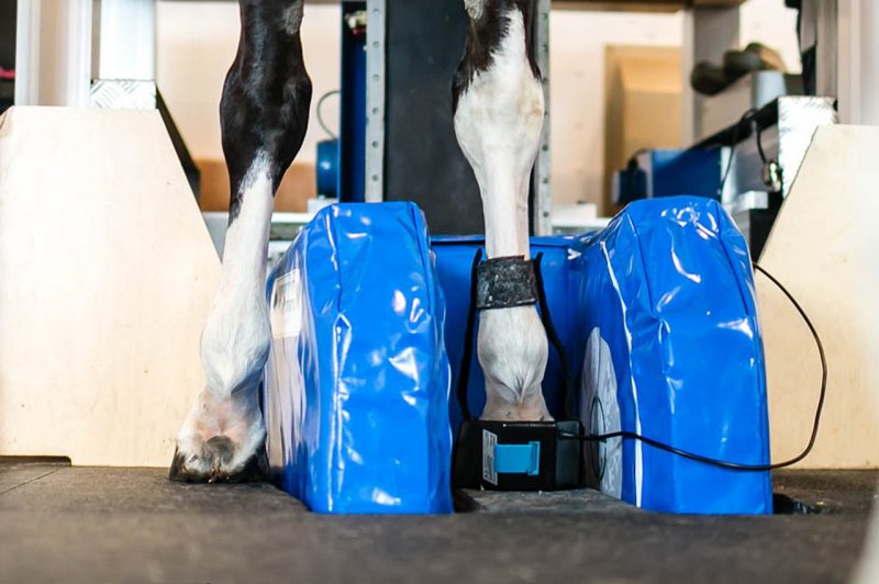
The Equine Medical Center offers low-field magnetic resonance imaging (MRI) services for evaluation of the lower limbs in the standing, sedated horse. With the launch of our MRI in 2004, we became the first equine hospital on the East Coast to offer this service.
Benefits of MRI
- Magnetic resonance imaging provides detailed images of bones and soft tissues in the lower limbs and can identify areas of inflammation and changes that cannot be detected by radiographs and ultrasound.
- MRI is recommended for identifying the source of pain to ensure an accurate diagnosis when evaluating the foot or lower limb.
Learn more about the procedure
-
Article Item
Appointments and referrals:
703-771-6800
To schedule an appointment, refer a patient, or inquire about diagnostic imaging or other clinical services, please call 703-771-6800 or email emcinfo@vt.edu.